|
FETAL HYDROCELE |
ETIOLOGY
|
- Normal appearance in the third trimester male fetus, as fluid frequently accumulates within the patent processus vaginalis during descent of the testis and epididymis. A small stable hydrocele is probably normal and such fluid is usually absorbed by one year of age.
- Fetal ascites or hydrops.
- Intrauterine intraperitoneal transfusion for non-immune hydrops.
PATHOGENESIS
|
- Between
the seventh week and birth, the testes descend into the scrotum due to
shortening of the gubernaculum. The testes pass through the inguinal canal
in the anterior abdominal wall.
- After
the 8th week, a peritoneal evagination, the processus
vaginalis, forms just anterior to the gubernaculum. It forms the inguinal
canal by pushing out sock-like extensions of the transversalis fascia, the
internal oblique muscle and external oblique muscle, The inguinal canal
extends from the base of the inverted transversalis fascia (the deep ring)
to the base of the everted external oblique muscle (the superficial ring).
After the processus vaginalis has evaginated into the scrotum, the
gubernaculum shortens and pulls the gonads through the canal. The gonads
always remain within the plane of the subserous fascia associated with the
posterior wall of the processus vaginalis.
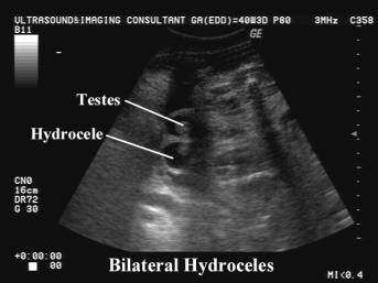
- By the end of the pregnancy the testes have completely entered the scrotal sac. The gubernaculum is reduced to a ligamentous band attaching the inferior pole of the testis to the scrotal floor. Within the first year after birth the superior part of the processus vaginalis is usually obliterated leaving a distal remnant sac, the tunica vaginalis, which lies anterior to the testis. Its lumen is normally collapsed but sometimes it may fill with serous secretions forming a testicular hydrocele.
ULTRASOUND
|
- Although literature suggests that testicular descent occurs between 18 and 32 weeks, hydroceles have been reported from 27 weeks until term.
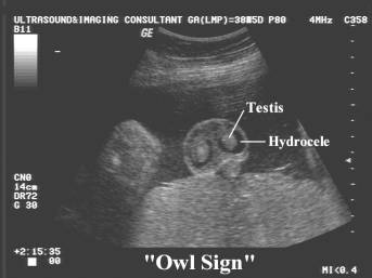
- "Owl sign" (1).
ASSOCIATIONS
|
- Fetal hydrops.
- Meconium peritonitis (2).
- Inflammation from Meckel diverticulum (3).
DIFFERENTIAL DIAGNOSIS
|
1. Testicular
torsion:
·
Mostly unilateral and
on the left side.
·
Acute phase - hypoechogenic testicle (due to edema) or a
heterogeneous pattern (due to necrosis), but a hydrocele can be an early sign
of a testicular torsion.
·
Chronic phase - a
small rounded, hypoechogenic area, with a peripheral echogenic ring due to
calcium deposits.
2. Inguinoscrotal
hernia - rare scrotal mass with multicystic structure. Peristaltic movements
can occasionally be observed.
PROGNOSIS
|
Hydroceles are a common antenatal finding that should be viewed as
physiologic as long as no other fetal abnormalities are detected (4).
REFERENCES
|
- Sherer DM, Smith SA. Suggested "Owl Eye Sign" in fetal sonography. J Ultrasound Med 1990;9:690.
- Ring KS, Axelrod SL, Burbige KA et.al. Mecomium hydrocele: An unusual etiology of a scrotal mass in the newborn. J Urol 1989;141:1172.
- Wright JE, Bhagwandeen SB. Antenatal perforation of Meckel's diverticulum presenting as an inflamed hydrocele. J Pediatr Surg 1986;21:989.
- Pretorius DH, Halsted MJ, Abels W et.al. Hydroceles detected prenatally: Common physiologic phenomenon. J Ultrasound Med 1998;17:49-52.