Ultrasound of Cleft Lip and Palate
|
|
Type
|
Chromosomal |
|
Type
1 |
O% / 20% |
|
Type
2 |
20% / 33% |
|
Type
3 |
30% / 50% |
|
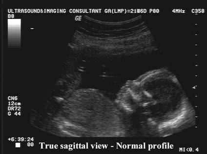
Type 4 sagittal views of the face. |
52% / 100% |
|
Type
5 |
0% / 100% |
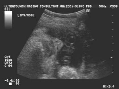
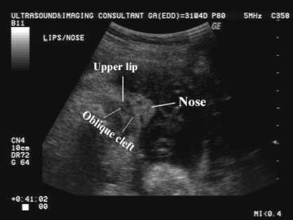
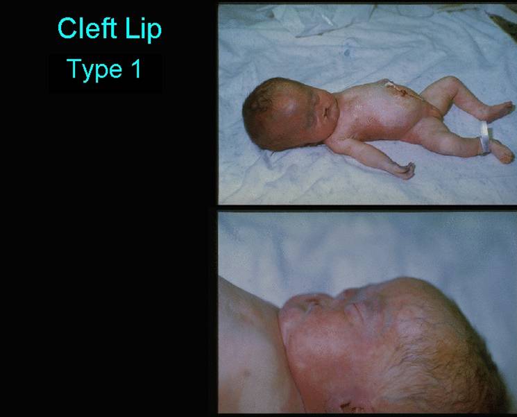
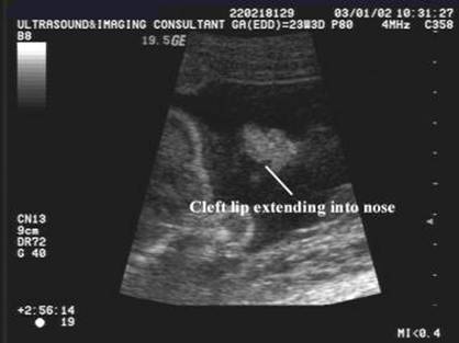
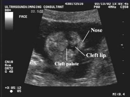
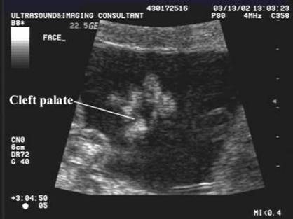
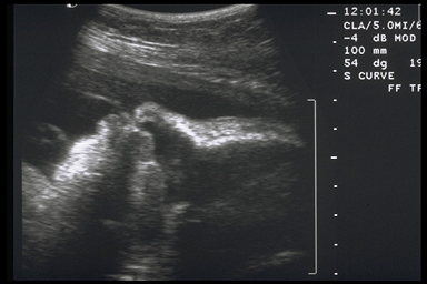
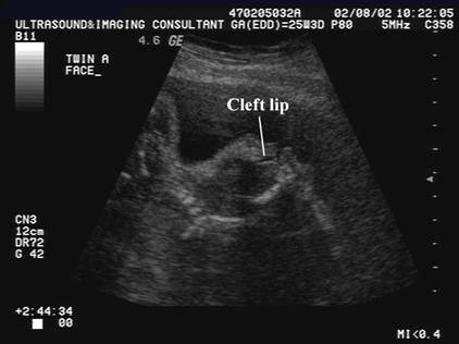
Type 1 Cleft
lip
|
|
|
|
|
|
|
|
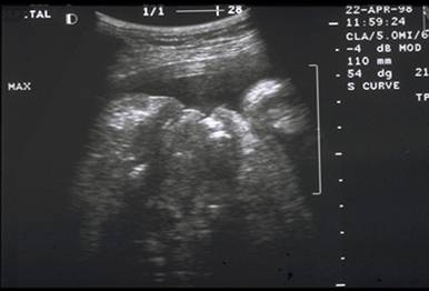
Cleft lip
and palate
|
|
|
|
|
|
|
|
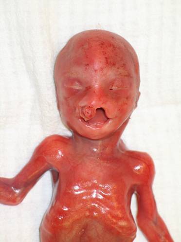
Cleft
lip and palate
|
|
|
|
|
|
|
|
|
|
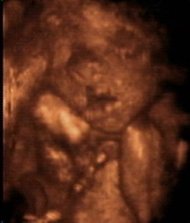
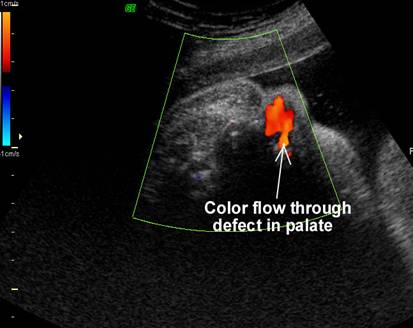
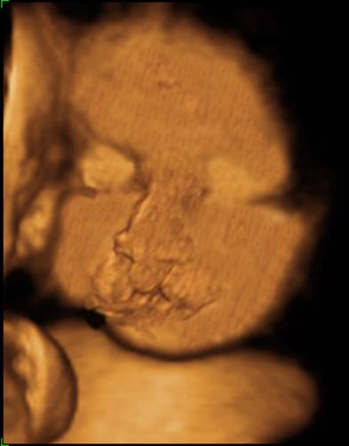
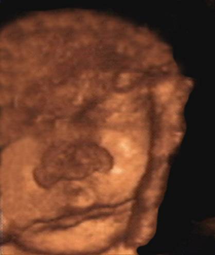
3D - Unilateral cleft lip
|
|
|
|
|
|
|
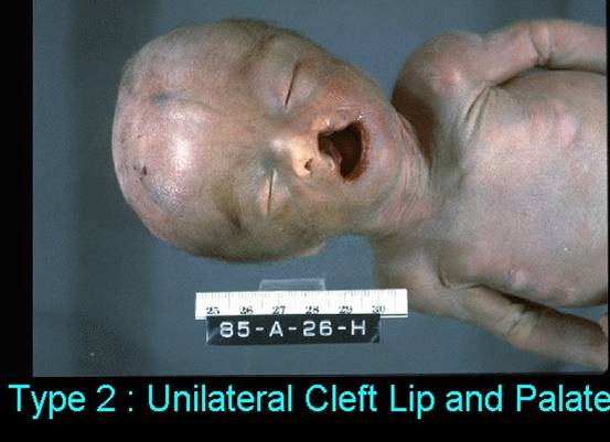
Type 2 – Cleft lip and palate
|
|
|
|
|
|
|
|
|
|
|
|
|
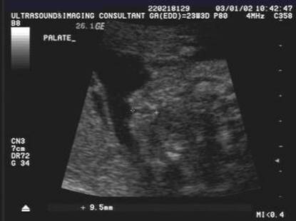
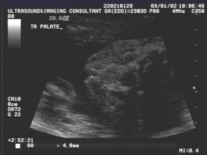
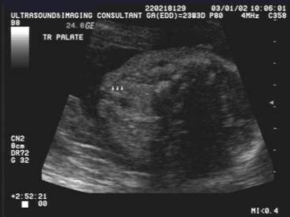
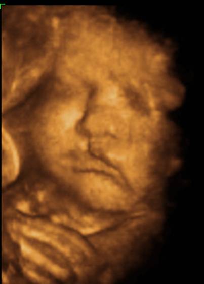
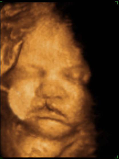
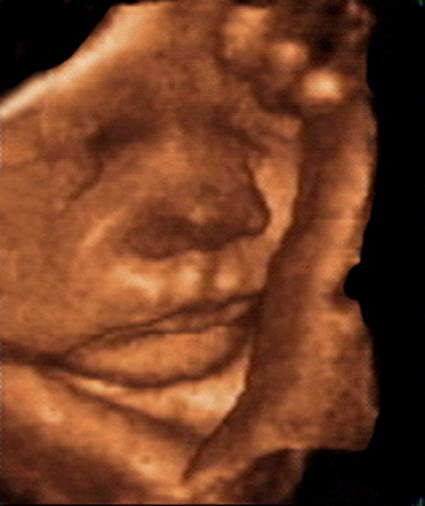
3D images |
|
Type
3 – Cleft lip and palate |
|
|
|
|
|
|
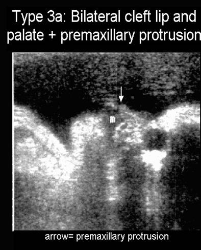
Bilateral cleft lip with pre-maxillary protrusion |
|
|
|
|
|
|
|
|
|
|
|
|
|
|
|
|
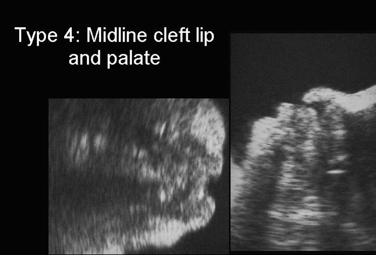
Type 4 |
|
|
|
|
|
Midface
hypoplasia |
|
|
|
|
|
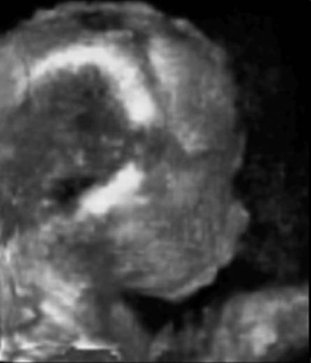
Type 5 |
|
|
|
|
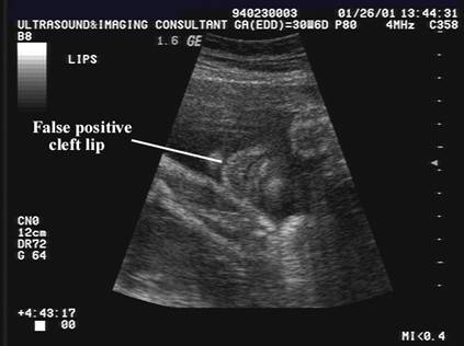
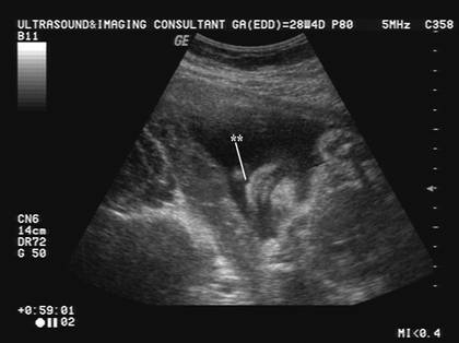
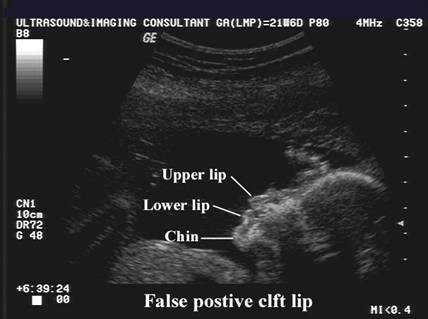
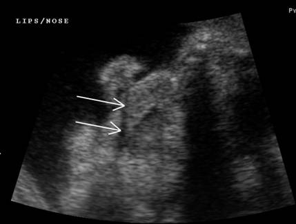
False
positive cleft lip
Normal indentation on upper lip - philtrum |
|
|
|
|
|
|
|
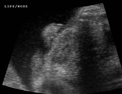
False positive cleft of upper lip
|
|
|
|
|
|
False
positive cleft lip ·
Artifactual
line across the upper lip disappears on changing transducer orientation. |
|
|
|
|
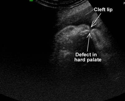
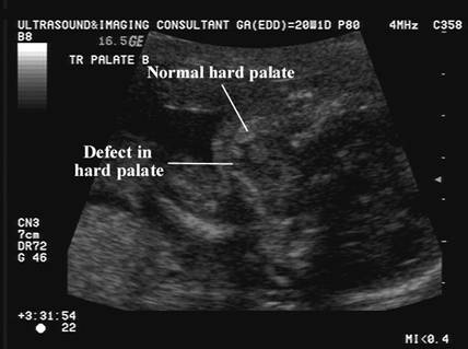
Cleft Lip and Palate
|
|
|
|
|
|
|
|
|
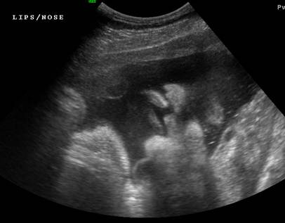
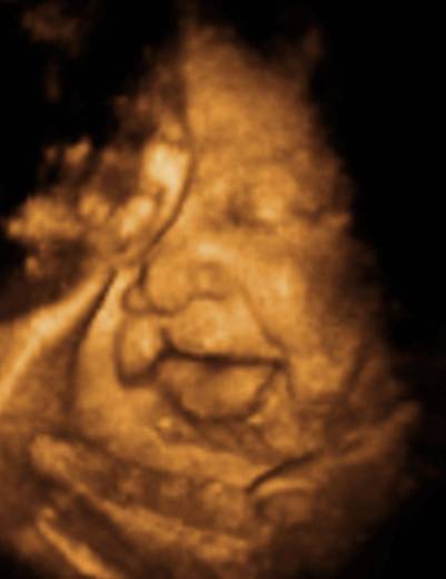
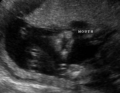
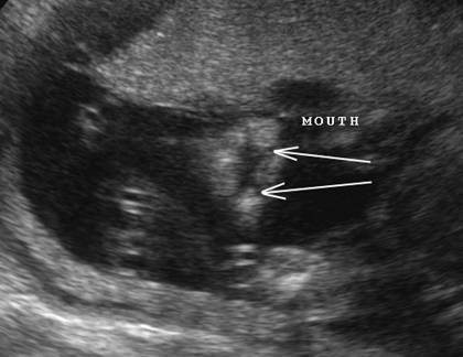
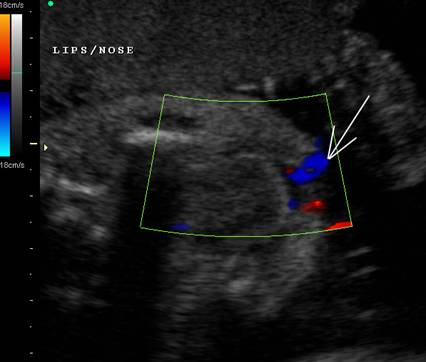
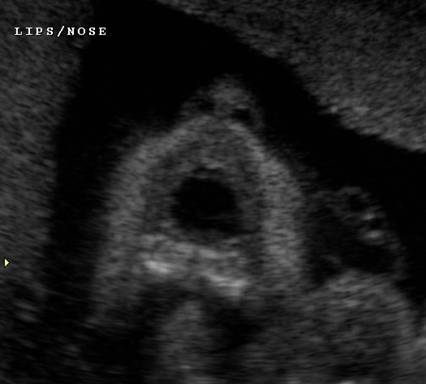
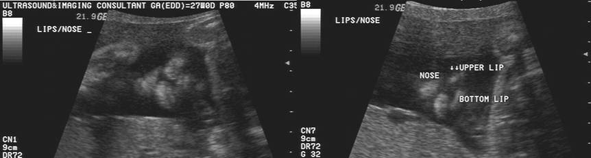
Fetus with mouth open demonstrates a defect
which is part of the open mouth. The lower two images demonstrates normal
upper and lower lips. |
|
|
|
|
|
|
|
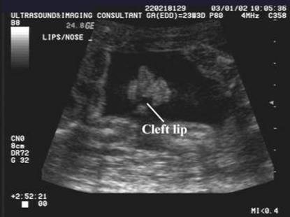
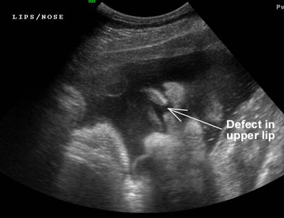
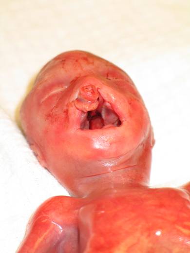
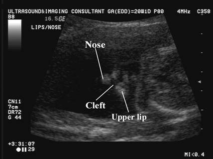
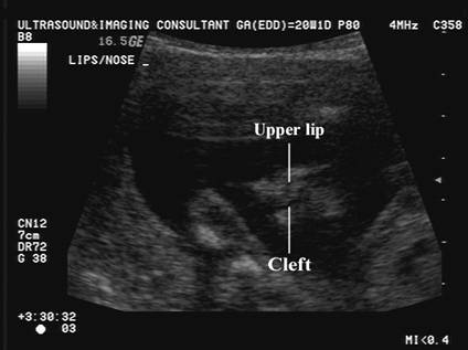
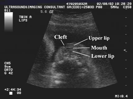
Cleft of upper lip
|
|
|
|
|
|
|
|
|
|
|
REFERENCES
|
- Nyberg DA, Sickler GK, Hegge FN et.al. Fetal cleft lip with and without cleft palate: