NUCHAL FOLD (EDEMA) |
Subcutaneous accumulation of fluid considered an early sign of fetal hydrops (1).
ULTRASOUND |
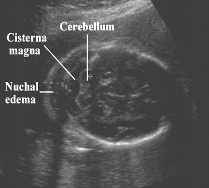
- Echogenic area between the skin surface and the occiput or spine, representing subcutaneous accumulation of fluid.
- Seen between 14 and 18 weeks gestational age (at 18-20 weeks measurement of nuchal thickness becomes unreliable due to the wide range in the normal rate of development of the fetal neck muscles and subcutaneous tissue).
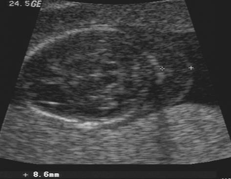
- Echogenic area greater than 6mm
|
|
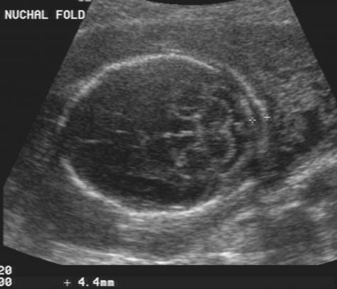
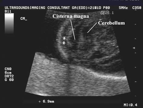
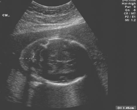
Calipers placed from outer skull table to outer skin surface Normal range =
1- <5 mm Borderline
range = 5 – 5.9 mm Abnormal = 6
mm |
|
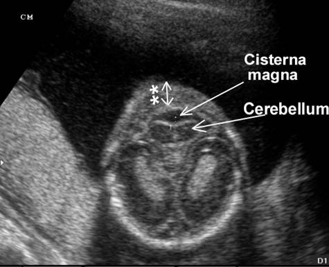
Measurement can be overestimated by angling caudally, intersecting the inferior level of the cerebellum and occiput |
|
|
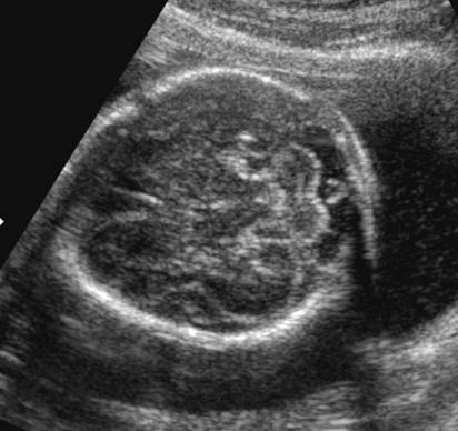
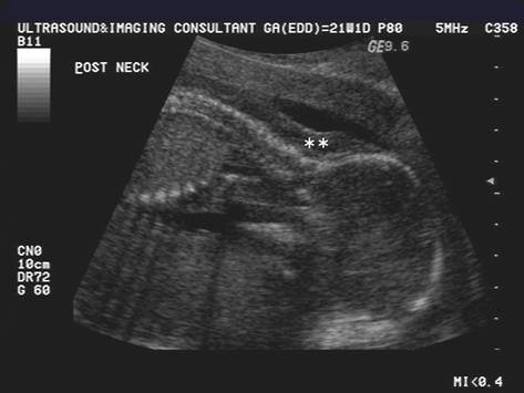
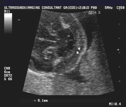
Nuchal thickening
(**) on the left image is created by a steep angle of insonation.
By changing the angle the nuchal soft tissues are
normal (right image). |
|
|
|
|
|
Measurement can be overestimated by angling caudally, intersecting the inferior level of the cerebellum and occiput |
|
|
|
|
|
|
|
|
Diffuse nuchal edema |
|
|
|
|
|
|
|
|
|
|
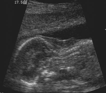
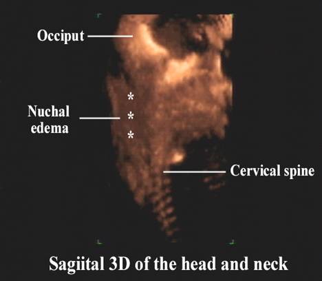
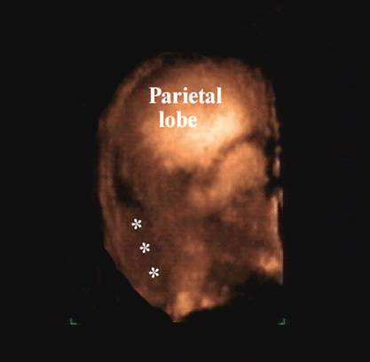
- Localized or diffuse.
- Ballotment of the fetal head is characteristic.
- Link to thick nuchal fold in Down syndrome
ETIOLOGY |
- Diverse causes including: trisomies, cardiovascular and pulmonary defects, skeletal dysplasia, congenital infection, metabolic and hematological disorders.
- Karyotyping is recommended in all cases (2).
- Nuchal
edema + no other structural abnormalities (cardiac or non cardiac
Þ about one third of fetuses will have a karyotypic abnormality (usually Down Syndrome and less commonly Trisomy 18)
Þ about two thirds will have a normal karyotype. - Nuchal
edema + cardiac and non-cardiac structural abnormality
Þ majority of fetuses will have chromosomal aneuploidy. - Nuchal thickening in Down syndrome may resolve prior to 16-18 weeks (3).
REFERENCES |
- Nicolaides KH, Azar G, Snijders RJM, Gosden CM. Fetal Nuchal Oedema: Associated Malformations and Chromosomal Defects Fetal Diagn Ther 1992, 7:123-131
- Suchet I. Ultrasonography of the fetal neck in the first and second trimesters. Part 2. Anomalies of the posterior nuchal region. Can Assoc Radiol J 1995; 46:344-352
- Bromley B, Benacerraf
- Comas C, Martinez JM, Ojuel J et.al. First trimester nuchal edema as a marker of aneuploidy. Ultrasound Obstet Gynecol 1995; 5:26-29