|
ULTRASOUND OF
MYELOMENINGOCELES |
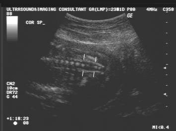
- Normal
Spine
- Myelomeningocele.
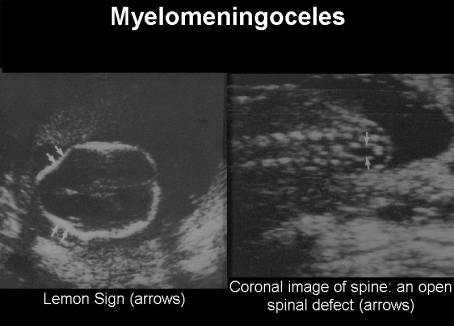
- Spinal lesions associated with open spinal; defect seen in only 80%-90% of scans performed at 18 weeks GA.
- Posterior transaxial
scan is the best plane for assessing the spine. The axial view is
the best view as the three ossification centers of the vertebra (one in
body and two in neural arch) and the soft tissues covering the spine are
readily visualized.
- The laminae fail to converge towards the midline and are divergent. The skin folds and subcutaneous tissues are well assessed, and are absent at the level of the defect.
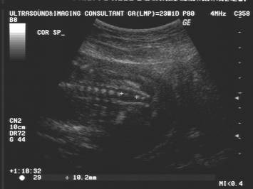
- Coronal image - Affected bony segment is divergent replacing the typical parallel lines of the normal arches. The interpedicular distance is usually increased, rather than the normal tapering towards the sacrum.
|
|
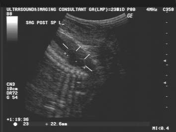
- Sagittal images - useful plane for determining both the level and extent of the lesion. S4 = most caudal ossification center visible in 2nd trimester while S5 is most caudal level in 3rd trimester. Can sometimes appear normal even though there is a large defect and therefore other imaging planes are essential.
- In cases of meningocele and myelomeningocele, the sac is best seen in the posterior longitudinal and posterior transaxial planes. It is important to get the spine away from the uterine wall.
- The spinal level can be accurately estimated by counting the vertebral segments:
- In 95% of cases S1 is at the top of the iliac wing.
- L5 and S2 are each at the top of the iliac wing in 2% of cases.
- A coronal view may be used to count down from the 12th rib and thus determine the level of the defect.
- By 22 weeks, S2 is ossified ion most fetuses, but a significant percentage of fetuses will have non-ossified or incomplete ossification of the neural arch centers in the sacrum at 23-24 weeks.
- One may also count up from the last ossified sacral segment (usually S4 in the second trimester and S5 in the third trimester).
- Studies have shown that the ossification pattern of the vertebral body is independent of that of the neural arches. In the lower spine, ossification of the neural arch centers occurs in a caudal direction at a rate of 1 vertebral level every 2-3 weeks after 16 weeks menstrual age.
- Evaluation of the conus medullaris can be obtained using a high frequency transducer when the fetus is in a prone position. Spinal cord determination can be accurately identified as early as 19 weeks. By the time the conus is visualized sonographically, it should be located within the normal adult range (L2-3 or higher).
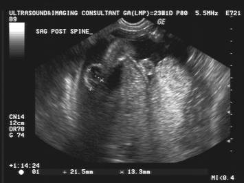
- It is important to estimate the size, location and contents of the sac.
- Evaluation of neurological deficit can be estimated by assessing flexion and extension of the lower limbs from the hips to the legs. It is also important to search for an associated club foot.
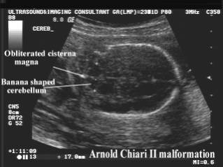
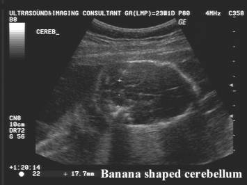
- Cranial
abnormalities associated with open spinal defect seen in 90%-100% of
scans at 18 weeks GA. A Chiari type II
malformation is almost always associated with a myelomeningocele. The
malformation is characterized by a small posterior fossa, with caudal
displacement of the inferior cerebellum and tonsils, pons, medulla and
fourth ventricle into the upper cervical canal.
|
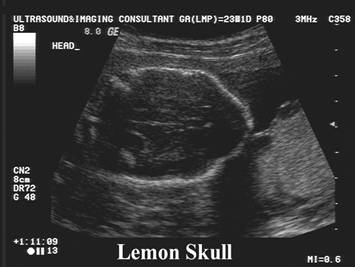
Frontal bone convacity
due to inward scalloping of the frontal bones of the calvarium such that the
head appears shaped like a lemon. It may be visualized when the spinal defect
is small and difficult to see.
|
|
|
|
|
|
|
|
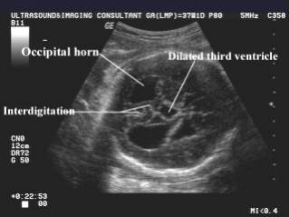
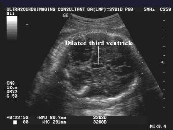
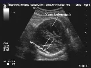
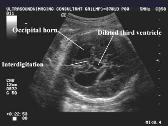
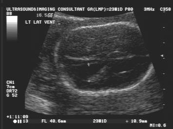
Ventriculomegaly due to stenosis, dilatation or shortening of the aqueduct of Sylvius
|
|
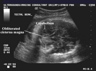
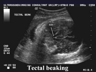
Tectal beaking – pointed appearance of the quadrigeminal plate
|
|
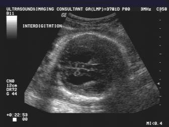
Interdigitation of gyri across the midline due to fenestration of the falx
|
|
|
|
|
Associated with callosal agenesis |
|
3. Location.
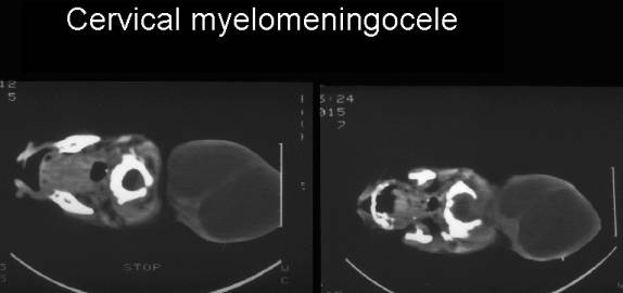
Cervical myelomeningocele |
|
|
|
|
|
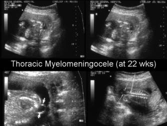
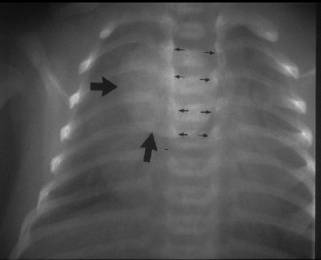
Thoracic myelomeningocele |
|
|
|
|
|
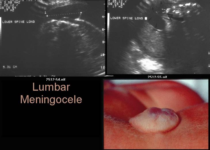
Lumbar meningocele |
|
|
|
|
|
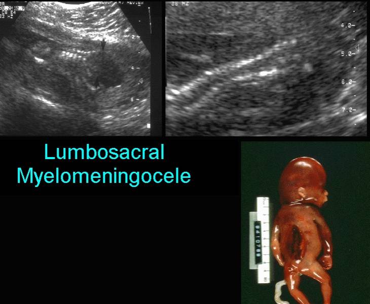
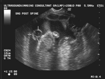
Lumbar myelomeningocele |
|
|
|
|
|
Lumbar myelomeningocele |
|
|
|
|
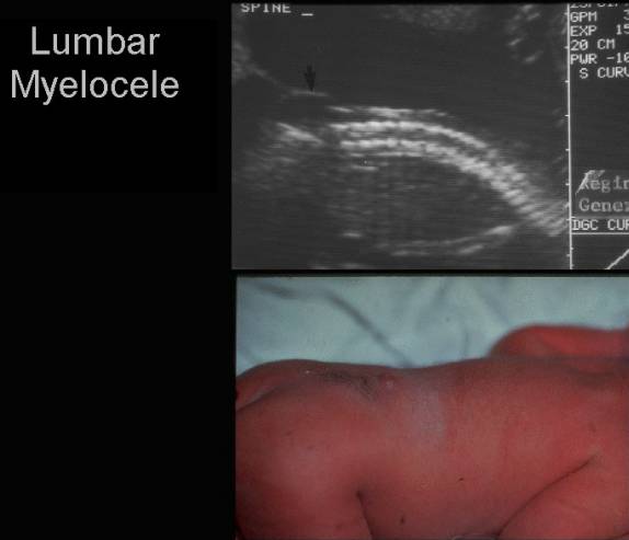
- Lumbar Myelocele.
|
|
|
|
Video clip of a Myelomeningocele
|
|
|
|
|
|