|
ULTRASOUND IN
LISSENCEPHALY |
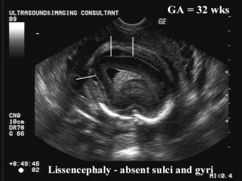
- Without a previous history of an affected child the diagnosis cannot be reliable made until 26-28 weeks (when sulci and gyri become well defined sonographically).
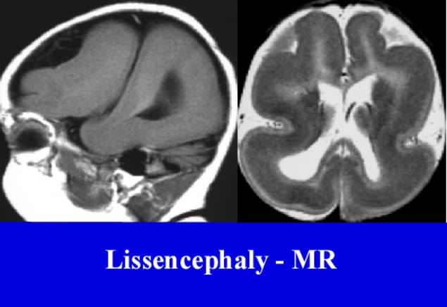
- Absent gyri and a smooth brain.
- A
detailed knowledge of the gestational age at which the cerebral sulci
become visible is essential.
|
|
|
|
|
|
|
|
|
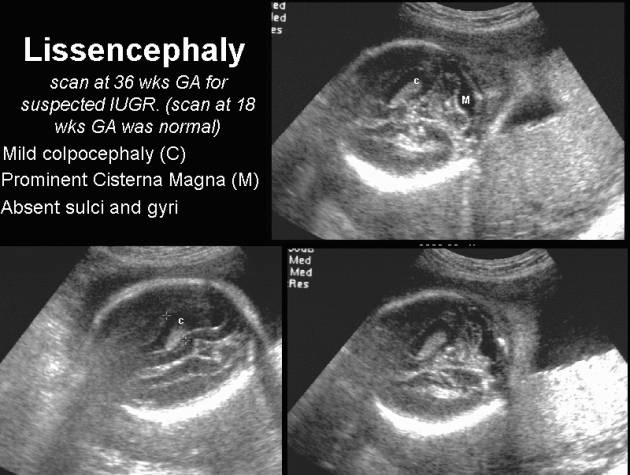
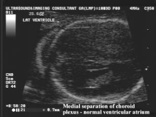
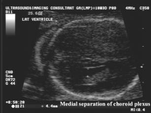
- Mild dilatation of the lateral ventricles (with colpocephaly).
- Rarely marked hydrocephalus.
- Microcephaly.
- Hypoplasia or agenesis of the corpus callosum.
- Dandy-Walker malformation (50%).
- Posterior cephalocele (25-50%).
- Polyhydramnios (impaired fetal swallowing).
- IUGR (50%).
- Less common abnormalities that
have been reported include:
micromelia, polydactyly, club foot, omphalocele, duodenal atresia, hepatosplenomegaly, cardiac and renal anomalies.
|
|
|
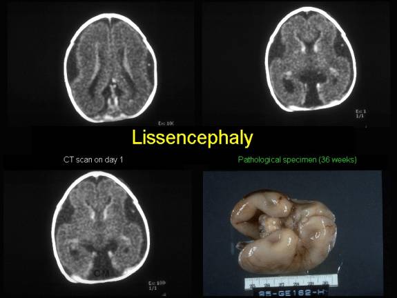
Lissencephaly (CT and Pathological specimen):
|
REFERENCES
|
- Saltzman DH, Krauss CM, Goldman JM et.al. Prenatal diagnosis of lissencephaly. Prenat Diagn 1991;11:139-143.
- Okamura K, Murotsuki J, Sakai T et.al. Prenatal diagnosis of lissencephaly by magnetic resonance imaging. Fetal Diagn Ther 1993;8:56-59.