|
DILATATION OF THE
FETAL URINARY TRACT |
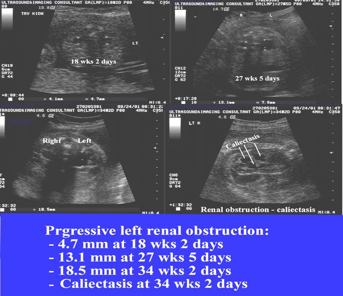
Prenatal detection of renal dilatation has led to earlier postnatal diagnosis of significant renal obstruction. This has resulted in better postnatal monitoring and early surgical intervention if necessary. Renal obstruction in the newborn is often silent (less than 25% of infants are diagnosed within the first year of life) (1), however antenatal scans have a sensitivity of 90-100% in detecting obstructive uropathy (2-5). Depending on the diagnostic criteria that are applied, about 1% of all fetuses may demonstrate a structural abnormality of the genitourinary tract (6).
Urinary tract distention is also influenced by maternal hydration (7,8), and
the time of the scan. Persutte et.al. have demonstrated that the size of the
fetal renal collecting system is highly variable over a 2 hour period (9). 70%
of their cases (14/20) had both normal (<4 mm) AP diameter and abnormal (4
mm or above) values during the 2 hour period. Implications based on one single
measurement should be viewed with caution.
DEFINITIONS |
|
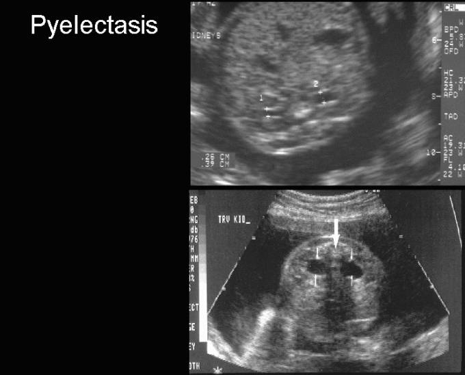
Pyelectasis = Increased AP diameter of the renal pelvis. |
|
|
|
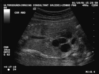
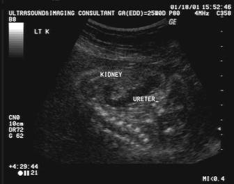
Dilatation of
the ureter. Multiple communicating cystic
lesions represent a dilated ureter on the sagittal
image.demonstrates communication between the cyst and the renal pelvis. |
|
|
|
|
|
Sensitivity and odds of detecting an obstructed kidney in relation to renal pelvis diameter. |
|
Relationship between fetal renal pyelectasis and chromosomal aneuploidy. |
REFERENCES |
- Snyder HM, Lebowitz RL, Coldny AH et.al. Ureteropelvic junction obstruction in children. Urol Clin North An 1980;7:273-289.
- Chitty LS, Hunt GH, Moore L et.al. Effectiveness of routine ultrasonography in detecting fetal anomalies in a low risk population. Br Med J 1991;303:1165-1169.
- Corteville JE, Gray DL, Crane JP. Congenital hydronephrosis: correlation of fetal ultrasonic findings with infant outcome. Am J Obstet Gynecol 1991;165:384-388.
- Helin I, Persson PH. Prenatal diagnosis of urinary tract abnormalities by ultrasound. Pediatrics 1986;78:879-883.
- Paduano L, Goglio L, Bembi B et.al. Clinical outcome of fetal uropathy. 2. Sensitivity of echography for prenatal detection of obstructive pathology. J Urol 1991;146:1097-1098.
- Goncalves LF, Jeanty P, Piper JM et.al. The accuracy of prenatal ultrasonography in detecting congenital abnormalities. Am J Obstet Gynecol 1994;171:1606-1612.
- Babcock CJ, Silvera M, Drake C et.al. Effects of maternal hydration on mild pyelectasis. J Ultrasound Med 1998;17:539-544.
- Robinson JN, Tice K, Kolm P et.al. Effect of hydration on fetal renal pyelectasis. J Ultrasound Med 1998;92:137-141.
- Persutte WH, Hussey M, Chyu J et.al. Striking findings concerning the variability in the measurement of the fetal renal collecting system. Ultrasound Obstet Gynecol 2000;15:186-190.
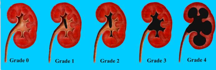
- Fernbach SK, Maizels M, Conway JJ. Ultrasound grading of hydronephrosis: introduction to the system used by the Society for fetal urology. Pediatr Urol 1993;23:478-480.