|
ULTRASOUND DIAGNOSIS
OF IUGR |
- Fetal morphometric
indices.
- Fetal age determination.
- BPD, HC.
- AC.
- Femur length.
- Humerus, ulna, radius, tibia,
fibula (Tables / Graphs)
- Normal Transcerebellar Diameter (Table / Graph)
- Ratio of HC/AC.
- Fetal mass determination.
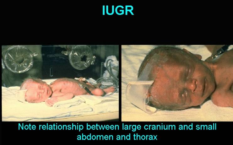
- Fetal proportions (symmetrical versus asymmetrical).
- Amniotic
fluid volume.
- Fetal
fat distribution.
- Fetal
biophysical profile scoring.
- Placental
assessment.
- Doppler blood flow velocity waveforms.
- Categories based on estimated fetal weight and umbilical artery S/D ratio (1)
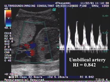
- Umbilical artery RI/PI
- Increases.
- Descending aorta RI/PI
- Increases.
1. Group 1: AGA fetuses with normal S/D ratio.
- Renal artery RI/PI -
Increases.
2. Group 2: AGA fetuses with abnormal S/D ratio (above 90th
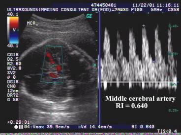
- Cerebral arteries
RI/PI - Decreases. centile
90th centile for GA). Increased neonatal morbidity.
- Adrenal artery RI/PI -
Decreases.
3. Group 3: SGA fetuses
with normal doppler studies - Outcome
- Coronary artery RI/PI
- Decreases.
the same as AGA cohorts
- Splenic artery RI/PI -
Decreases.
5. Group 4: SGA fetuses with abnormal doppler studies –.
- Peripheral pulmonary artery RI/PI - Increases. Increased perinatal mortality
- Fetal compensation:
- “Brain
sparing effect”.
|
IUGR with
fetal compensation (“brain-sparing” effect) Middle cerebral artery / Umbilical
artery Ratio = 0.64 / 0.84
= 0.76 Normal = > 1 |
|
|
|
|
|
|
|
·
Increased blood flow to myocardium (dilated
coronary arteries)
|
|
Video clip of IUGR
- Centralization of fetal blood flow Video clip of IUGR
- Absent end-diastolic flow in the umbilical artery
|
|
|
|
|
|

REFERENCES |
1. Ott WJ. Intrauterine growth restriction and doppler ultrasonography. J Ultrasound Med 2000;19:661-665.