|
VOLUME DISPLAY |
Three-dimensional Doppler is an area of intense research using both power Doppler and traditional color flow. One particular area of interest has been the detection of abnormal placental adherence (accreta, inccreta, or perccreta) with or without bladder involvement. Three-dimensional color Doppler has allowed the investigators to visualize all three orthogonal planes of the placental myometral unit, a technique which appears to be complimentary to standard 2D sonography in clarifying abnormal neovascularization in patients with placenta accreta.(51, 52) Power Doppler in 3D is currently being studied as a way to visualize the fetal vascular system for prenatal diagnosis of anomalies, although more information is required to determine the utility of these techniques above and beyond the 2D Doppler.(53). Real-time capabilities in the use of Doppler will be necessary in 3D applications with similar frame-rates as those available today in 2D Doppler examinations.
TYPES |
|
Multiplanar reconstruction (MPR) |
|
Surface rendered |
|
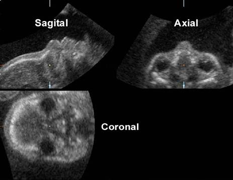
Skeletal view |
|
Angio view (3D Doppler) |
|
Glass Body rendering |
MULTIPLANAR RECONSTRUCTION |
SURFACE RENDERED |
|
Some Examples |
|
|
|
|
|
|
|
|
|
|
|
|
|
|
|
SKELETAL VIEW |
|
Examples |
|
|
|
ANGIO / 3D DOPPLER |
Three-dimensional Doppler has been investigated using both power Doppler and traditional color flow. One particular area of interest has been the detection of abnormal placental adherence (accreta, inccreta, or perccreta) with or without bladder involvement. Three-dimensional color Doppler has allowed the investigators to visualize all three orthogonal planes of the placental myometral unit, a technique which appears to be complimentary to standard 2D sonography in clarifying abnormal neovascularization in patients with placenta accrete (1,2).
|
Example |
1.
Hull AD,
2. Chou MM, Tseng JJ, Ho ESC, Hwang JI. Three-dimensional color power Doppler imaging in the assessment of uteroplacental neovascularization in placenta previa increta/percreta. Am J Obstet Gynecol 185(5):1257-1260, 2001.
GLASS BODY |
|
Example |
INVERSION MODE
|
The “inversion mode” is a new rendering algorithm that transforms echolucent structures into echogenic voxels. Thus, anechogenic structures such as the heart chambers, lumen of the great vessels, stomach and bladder appear echogenic on the rendered image, whereas structures that are normally echogenic prior to gray-scale inversion become anechoic (1,2).
The
“inversion mode” can be used at the time of scanning or after the
acquisition of volume datasets with both 3D and 4D techniques. This rendering
mode provides more anatomical detail than other rendering algorithms, such as
the transparent mode, in the evaluation of hollow anatomical structures (2).
The quality of the image rendered with the ![]() inversion mode
inversion mode![]() depends on
the quality of the volume dataset acquired. In a study by Espinosa and co-worhers (3) it was shown that the best results were
obtained by a combination of
depends on
the quality of the volume dataset acquired. In a study by Espinosa and co-worhers (3) it was shown that the best results were
obtained by a combination of ![]() surface smooth
surface smooth![]() and
and ![]() gradient light
gradient light![]() filters.
filters.
The
3D/4D “inversion mode” rendering algorithm can improve the prenatal
visualization of a dilated azygos or hemiazygos vein and their spatial relationships with the
surrounding cardiovascular structures (2). The “inversion mode”
algorithm improves prenatal visualization of both dilated azygos
and hemiazygos veins, as well as their spatial
relationships with the surrounding vascular structures
- Goncalves LF, Espinoza J, Lee W et.al. Three- and four-dimensional reconstruction of the aortic and ductal arches using inversion mode: a new rendering algorithm for visualization of fluid-filled anatomical structures. Ultrasound Obstet Gynecol 2004; 24: 696-698
- Lee W, Goncalves LF, Espinoza J, Romero R. Inversion mode: a volume analysis tool for three-dimensional sonography. J Ultrasound Med 2005; 24: 201-207
- Espinoza J, Gonçalves LF, Lee
W, Mazor M et.al. A novel method to improve prenatal
diagnosis of abnormal systemic venous connections using three- and
four-dimensional ultrasonography and
 inversion
mode
inversion
mode Ultrasound Obstet Gynecol
2005;25:428-434.
Ultrasound Obstet Gynecol
2005;25:428-434.
VOLUME MEASUREMENTS |
Another important clinical application of 3D is volume measurements calculations based on 3D volume acquisition.
- Brummer et al. demonstrated the accuracy of volume based three-dimensional sonography measurement on follicle aspiration performed using transvaginal needle guided technique (1).
- Other practitioners have also shown that three-dimensional sonographic methods provide accurate volume measurements of both regular and irregular objects thus providing an improvement, both in accuracy and examination time, over estimations using multiple 2D images (2,3).
- Specific area of study where volume measurements are clinically applicable include:
- serial fetal lung volume measurements for the prenatal detection of pulmonary hypoplasia.(4-6).
- volume measurements of the fetal thoraco-lumbar spine as well as kidneys, liver and heart have been established, and fetal liver volumes in normally grown fetuses have been compared to those small-for-gestational-age.(7-12)
- fetal brain volumes have been calculated using 3D scans with excellent intra and inter observer variability correlating well with other standard biometry at different gestational ages.(13)
- fetal weight estimates as well as growth parameters and improve the accuracy with which we can predict small-for-gestational-age infants. Fractional limb volume has also been investigated as an additional parameter to further improve accuracy in predicting birth weight, although this remains to be seen in larger scale studies(14-16)
- volume
measurements of the fetal placenta using 3D sonography
has not been particularly helpful to date in predicting
small-for-gestational-age infants.(17)
- Brunner M, Obruca A, Bauer P, Feichtinger W,. Clinical application of volume estimation based on three-dimensional ultrasonography. Ultrasound Obstet Gynecol 1995:6:358-361.
- Riccabona M, Nelson TR, Pretorius DH, Davidson TE. Distance and volume measurement using three-dimensional ultrasonography. J Ultrasound Med 1995;14:881-886.
- Riccabona M, Nelson TR, Pretorius DH. Three-dimensional ultrasound: accuracy of distance and volume measurements. Ultrasound Obstet Gynecol 1996;7:429-434.
- Bahmaie A, Hughes SW,
- Laudy JAM . Janssen MMM, Struyk PC, Stijnen T, Wladimiroff JW. Three-dimensional ultrasonography of normal fetal lung volume: a preliminary study. Ultrasound Obstet Gynecol 1998;11:13-16.
- Pohls UG, Rempen A. Fetal lung volumetry by three-dimensional ultrasound. Ultrasound Obstet Gyncol 1998;11:6-12.
- Schild RL, Wallny T, Fimmers R, Hansmann M. The size of the fetal thoracolumbar spine: a three-dimensional ultrasound study. Ultrasound Obstet Gynecol 2000;16:468-472.
- Schild RL, Wallny T, Fimmers R, Hansmann M. Fetal lumbar spine volumetry by three-dimensional ultrasound. Ultrasound Obstet Gynecol 13:335-339, 1999. Hsieh YY, Chang CC, Lee CC, Tsai HD. Fetal renal volume assessment by three-dimensional ultrasonography. Am J Obstet Gynecol 2000;182(2):377-379.
- Laudy JAM, Janssen MMM, Struyk PC, Stijnen T, Wallenburg HCS, Wladimiroff JW. Fetal liver volume measurement by three-dimensional ultrasonography: a preliminary study. Ultrasound Obstet Gynecol 1998;12:93-96.
- Kuno A, Hayashi Y, Akiyama M, Hata T, Yamashiro C, Yanaka H,Yanagihara T, Hata T. Three-dimensional sonographic measurement of liver volume in the small-for-
Dates. J Ultrasound Med 2002;21:361-366.
- Chang FM, Hsu KF, Ko
HC,
Gynecol 1997;9:42-48.
12. Endres LK, Cohen L. Reliability and validity of three-dimensional fetal brain volumes. J Ultrasound Med 2001;20:1265-1269.
13. Lee W, Deter RL, Ebersole JD, Huang R, Blanckaert K, Romero R. Birth weight prediction by three-dimensional ultrasonography. J Ultrasound Med 2001;20:1283-1292.
14. Song TB, Moore TR, Lee JY, Kim YH, Kim EK,. Fetal weight prediction by thigh volume measurement with three-dimensional ultrasonography. Obstets & Gynecol 2000;96(2):157-161.
15. Chang
FM,
16. Hafner E, Philipp K, Schuchter K, Dillinger-Paller B, Philipp T, Bauer P. Second-trimester measurements of placental volume by three-dimensional ultrasound to predict small-for- gestational-age infants. Ultrasound Obstet Gynecol 1998;12:97-102.